Research:Super-resolution fluorescence imaging of carbon nanotubes using a nonlinear excitonic process
When multiple excitons exist in a carbon nanotube, an exciton can scatter with another exciton and decay nonradiatively (this process is called exciton-exciton annihilation (EEA). Confinement and diffusion of the excitons in a nanotube lead to efficient EEA process upon collision with one another, resulting in a peculiar cubic dependence of the EEA rate on the density of excitons. The efficient EEA can be, for example, utilized in room-temperature single photon generation. As advanced techniques of super-resolution imaging often rely on the nonlinear optical response in fluorescence agents, the EEA process could play a key role in the development of nanotube-based biological imaging as well.
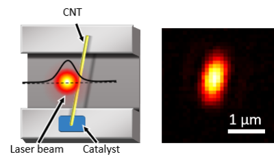
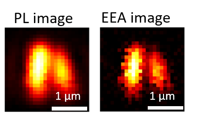
Here we demonstrate subdiffraction imaging of air-suspended CNTs by extracting the nonlinear EEA component using a typical confocal microscopy system. By combining two fluorescence images obtained at different excitation powers, an EEA rate image with enhanced resolution can be constructed.
In a confocal microscopy system, either a focused laser beam or a sample is scanned to form images. The width of the nanotube photoluminescence image is predominantly determined by the excitation laser beam profile, and thus much larger than the actual nanotube diameter of ~1.0 nm. If we approximate the laser beam profile by a Gaussian function exp(-2x2/r2) with r being the laser 1/e2 radius, the intensity profile of signals that have gα dependence becomes α1/2 smaller than the laser beam profile, where α is the power exponent of the generation rate dependence.
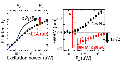
We can utilize the nonlinear power dependence on the generation rate to achieve enhanced spatial resolution. We extract the EEA component from photoluminescence intensities measured at two different excitation powers by subtracting the emission intensity at P1 (blue square) from that expected in the absence of EEA (green square). With the lower excitation power P2 fixed at 0.05 μW, we compare the width of photoluminescence images and exciton-exciton annihilation images at various P1 values, and find that the resolution enhancement through the EEA extraction is approximately 21/2. It is noteworthy that the EEA-derived nonlinearity in nanotubes enables subdiffraction imaging at a power density as low as ~300 W/cm2 with a continuous wave laser, whereas considerably higher intensities are employed for other subdiffraction techniques using conventional dyes.
The resolution enhancement by a factor of 21/2 may seem to contradict with the cubic dependence expected for EEA, but it is also confirmed by Monte Carlo simulations. Two different regimes hidden in the EEA rate dependence on exciton density are the keys to understanding this result.
To learn more about this work, please refer to:
Super-resolution fluorescence imaging of carbon nanotubes using a nonlinear excitonic process
Opt. Express
27, 17463 (2019).
